
 Neurobiology
Exploring the development of the neocortex
The neocortex is one of the most distinctive features of the human brain. Now we know, gene by gene, how its fourth layer develops.
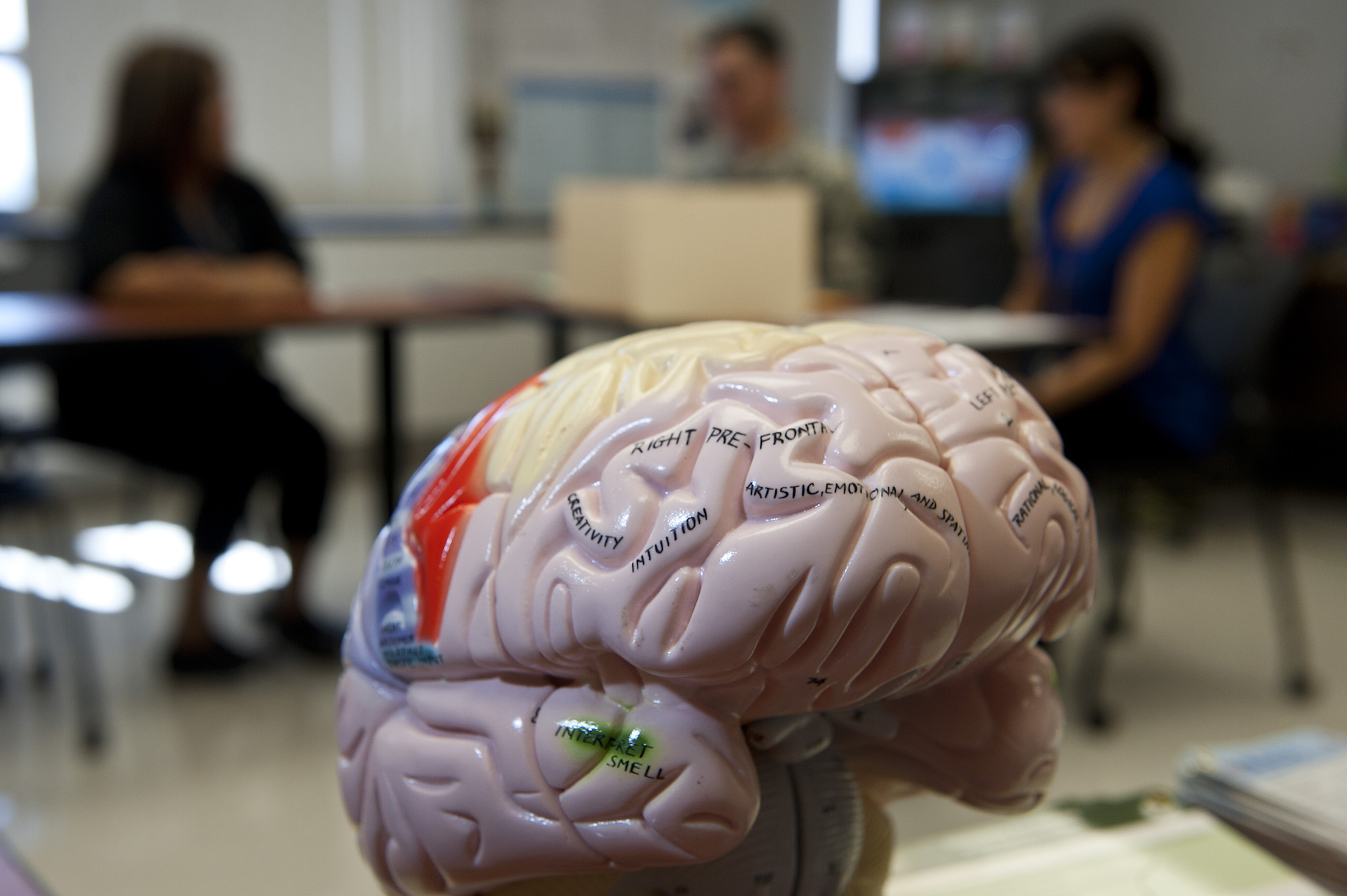
For all our life we've always wondered: what makes us human? A possible answer to that today would be: the neocortex. This evolutionary youngest part of our brain is a nest for such precious mental features as: emotions, reasoning, attention, communication etc. It's true that we are not the only ones who have a necortex: all other mammals have one too. But it seems that we've mastered our neocortex to very lofty heights.
So puzzling is this structure that we have a hard time understanding how it works. And when scientists do not understand how something works, they try to break it down into analytical concepts. That's how we've come to study the spatial and genetic architecture of the neocortex. How do neocortex neurons get their identity? How do they arrange connections with other neurons?
To understand that, we take help from our distant relatives - mice. Since mice have the neocortex too, we can observe the steps of its formation during animal development and try to get answers to our questions.
A distinctive feature of the neocortex is its layered structure: like a layered cake, it consists of different juxtaposed sets of neurons that form local and long-range circuits. Any cake has a base, and so does the neocortex. Its base is called the ventricular zone (VZ), a place where progenitor cells are located. Unlike embryonic cells, progenitor cells can divide only a limited number of times. During these divisions, one of the daughter cells remains a progenitor whereas the other becomes a neuron. The goal of each of these newborn neurons is pretty serious: they have to migrate from the ventricular zone to their place in one of the neurocortical layers, and form the right connections in order to correctly transmit signals after the animal is born. This is how newborn neurons become neocortical. The whole process is tightly controlled. But there is large number of brain disorders that result from malfunction of neuronal migration.
If you see a brain with the naked eye, you will see some slushy grayish mass, without being able to distinguish anything there. And even if you zoom in with a microscope, all neurons will look the same. To distinguish developing neurons of the neocortex from neurons that form other parts of the brain, Denis Jaubadon and his team, scientists from the Geneva University Medical Center (CMU), injected a fluorescent dye into the developing ventricular zone of a mouse. This allowed the scientists to visualize in a time-specific manner how the newborn neurons migrate towards a particular layer of the neocortex, because they shone green! But this was just the beginning.
Because fluorescence makes them readily identifiable, neurons that are migrating can be captured at certain moments and be genetically scrutinized. In other words, we can "look" inside the cells and see which genes are being expressed at the moment. Expression means that the genetic "blueprint" that is encoded in DNA is getting copied into a mobile molecule of RNA that will be further "translated" into a functional protein by special molecular machines. Differentially expressed genes are the basis of a cell's identity, because cells need different proteins to perform their tissue-specific functions.
So, scientists analyzed newborn neurons at different moments in their migration. What they got is information about how levels of expression of different genes change over time, while neurons are migrating. But there are dozens and dozens of such genes, and it's impossible to analyze them one by one. Therefore, the researchers used a machine-learning algorithm to sort this huge amount of data into four different classes, correspondent to the migration moments. This was possible to do because some single genes have been already associated with different migration stages of newborn neurons in studies of other research groups.
What could they learn from these data then? First, they observed that the genes in the developing neurons are not expressed in a random way. While newborn neurons migrate towards their layer, there's something interesting happening in them. Fixed groups of genes activate the expression of one another, creating a domino effect. It is like waves that initiate each other in a sequential order. And, for example, artificial expression of one gene in a wrong stage initiates a wrong wave and thus messes up migration and specification.
This study was focused on development of only one neocortical layer, the fourth one. So now we know, gene by gene, how this layer develops. One can say we have an atlas, a road map. Such an atlas opens the possibility for reverse engineering a directed specification of cortical neurons. This would be a tool to think up new testable hypotheses and concepts. Thus step-by-step we will break down the principles of how the neocortex functions. And that can have clinical applications: for when we understand the functions, we can seek ways to cure malfunctions.
Original Article:
L. Telley et al., Sequential transcriptional waves direct the differentiation of newborn neurons in the mouse neocortex. Science 351, 1443-1446 (2016)Edited by:
Dr. Carlos Javier Rivera-Rivera , Managing Editor
We thought you might like
How to print a brain - the initial steps
Nov 8, 2016 in Maths, Physics & Chemistry | 3.5 min read by Nieves CuboHacking the tryptophan metabolic process to reduce neurodegeneration
Apr 25, 2017 in Health & Physiology | 3 min read by Carlo BredaThe power of our adaptive immunity against Alzheimer’s Disease
May 10, 2017 in Health & Physiology | 3 min read by Daniele GuidoMore from Neurobiology
New, smaller-than-ever devices to help us understand how our brain works from the inside
Nov 8, 2024 in Neurobiology | 4 min read by Filippo DonatiCan we use a magnet to see brain inflammation?
Sep 25, 2023 in Neurobiology | 4 min read by Raquel Garcia-Hernandez , Santiago Canals , Silvia de SantisSurprising Behavior Changes in Genetically Modified Syrian Hamsters
Aug 30, 2023 in Neurobiology | 4 min read by Susan Lee , Kim Huhman , Jack TaylorTo achieve goals, we definitively need our neurons
Mar 10, 2023 in Neurobiology | 3.5 min read by Julien CourtinThe Impact of SARS-CoV-2 on the Brain: It Is All in Your Head
Feb 15, 2023 in Neurobiology | 3.5 min read by Meredith G. Mayer , Tracy FischerEditor's picks
Trending now
Popular topics