 Maths, Physics & Chemistry
How to print a brain - the initial steps

To understand the biological processes that take part within living organisms, and also as an alternative method to animal experimentation, researchers are trying to develop new models that mimic real tissues and organs, and that behave as the real ones. This field of research, tissue engineering, is a multidisciplinary field where medicine and engineering work together in order to replace, regenerate or improve a part or function of the body.
However, first approaches in vitro (in the lab, outside the normal biological context of the cells), were performed in 2D (two dimension, into a plane). These configurations did not allow cells to behave properly, as they do not recognize the environment where they are placed, which is far from being similar to their natural one.
3D printing is a technique that has been recently developed to produce three-dimensional objects. It works in the same way that a paper printer, but instead adding layers of ink to make words or images, 3D printers adds layers of a specific material to make an object. This technique has spread very much in recent years, reaching different fields such as fashion, engineering and medicine. Thanks to this spread and because of its layer-by-layer process, it has become a critical tool in tissue engineering. Due to the limitations of 2D tissue models, many research groups are focused on the development of new 3D tissue models using these machines. 3D printing tissue allows for the accurate positioning of cells, and also offers the opportunity of simultaneously printing several biomaterials. These materials will form the extracellular matrix (ECM) of the cells, which is the physical structure (scaffold) in which they will attach, proliferate and evolve until they organize in a tissue-like manner.
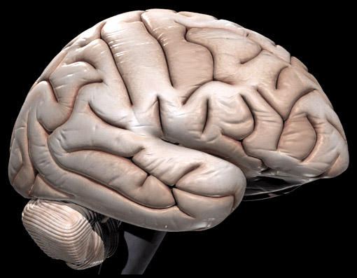
To understand this work, we have to know that the brain is an enormously complex organ structured into various regions of layered tissue, and has a lot of interconnected networks, which makes it especially difficult to replicate.
One of the research groups that is developing these 3D models was able to produce a new 3D layered brain-like structure using a hydrogel called "Gellan Gum" (GG) as the material for the scaffold. A modified peptide (short chain of amino acid) was added to the GG to improve its properties, and then they also added neural cells obtained from the brain of mice. These cells were covered by a thin layer of hydrogel before the 3D printing process, in order to protect them. Then, they used this mix (GG, peptide, and cells), to print a 3D model of a layered neural tissue, much like the brain!
Hydrogels, which are polymeric soft materials composed by a huge amount of water, have been adopted as standard extracellular matrix substitutes because cells can live in them. These materials are also very interesting because they can degrade as cells generate new tissue.
This group developed a material in which cells are able to live and thrive; and in addition, the modified peptide they added helped to promote cell adhesion and proliferation compared to the same materials without this specially modified peptide. They also studied other properties of the new material, such as the porosity and the diffusion of the material, which are important properties for facilitating nutrients, oxygen and waste exchange between cells and their culture medium. The results were also positive and they revealed the importance of different parameters carried out along the study that affects network size and its porosity. For example, if the pore is very small, cells cannot travel along the material, and the nutrients cannot be delivered. If they are very big, cells will not be comfortable and they could not create a new tissue
This first study was meant to validate a low-cost, simple extrusion technique and a bio-ink with the potential to organize neuron subtypes in layered structures, using a process that could be performed in any cell culture laboratory or bioprinter.
These brain-like structures offer the opportunity to provide a more accurate representation of 3D in vivo environments with applications ranging from cell behavior studies, understanding brain injuries and neurodegenerative diseases to drug testing. Finally, we cannot forget, that all these three-dimensional models offer an alternative to animal testing.
Original Article:
Lozano R, Stevens L, Thompson B et al. 3D printing of layered brain-like structures using peptide modified gellan gum substrates. Biomaterials. 2015;67:264-273. doi:10.1016/j.biomaterials.2015.07.022.Next read: The Lego bricks of the brain by Toshihiko Hosoya
Edited by:
Dr. Carlos Javier Rivera-Rivera , Managing Editor
We thought you might like
Hacking the tryptophan metabolic process to reduce neurodegeneration
Apr 25, 2017 in Health & Physiology | 3 min read by Carlo BredaThe power of our adaptive immunity against Alzheimer’s Disease
May 10, 2017 in Health & Physiology | 3 min read by Daniele GuidoToxic brain cells are a new target for treating neurodegeneration
May 24, 2017 in Health & Physiology | 3.5 min read by Shane A. LiddelowMore from Maths, Physics & Chemistry
Testing gravity through the distortion of time
Sep 20, 2024 in Maths, Physics & Chemistry | 3 min read by Sveva CastelloStacking molecular chips in multiple dimensions
Aug 30, 2024 in Maths, Physics & Chemistry | 3 min read by Lucía Gallego , Romain Jamagne , Michel RickhausReversible Anticoagulants: Inspired by Nature, Designed for Safety
Jun 12, 2024 in Maths, Physics & Chemistry | 4 min read by Millicent Dockerill , Nicolas WinssingerDistance-preserving moves always keep a point fixed
May 18, 2024 in Maths, Physics & Chemistry | 4 min read by Shaula FiorelliA resonance triggers chemical reactions between the coldest molecules
Apr 5, 2024 in Maths, Physics & Chemistry | 3 min read by Juliana Park , Wonyl ChoiEditor's picks
Trending now
Popular topics


